HTIC
HTIC
 Health Tech Lunch - 06.11.2019, 12h00 - Sierre, Techno-Pôle (Room Atlas)
Presentation by Traian Popa (EPFL)
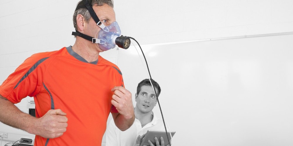
Title : Designing a network-based neuromodulation of the brain in health and disease
Abstract
Every part of the human brain is simultaneously involved in processing information from several different neuronal loops. A change in any part of this complex mosaic is going to tilt the equilibrium of many linked networks. For decades, both diagnostic and therapeutic approaches of brain stimulation have tapped one node at a time. This talk will review how multi-node approaches can open new perspectives into understanding the normal and pathological interaction within the brain.
Registration : https://doodle.com/poll/25bksby95cmefkhp
Presentation by Craig Shimasaki (Co-founder, ViRelieve) (full bio)
Title : Biotechnology Entrepreneurship: What You Need to Know to Start and Build a Successful Biotech Business in Valais
This lecture is for anyone who has dreamed that their technical or scientific ideas could one day become innovative products, but were unsure if this was even possible. This is for university, government and community leaders who want to see economic growth and diversity in their region. It is for all individuals who would like to work for a company that creates life-changing products through biotechnology entrepreneurship.
What we will cover :
Who should attend : Professors and students who have innovative discoveries and ideas. University and local government administrators who want to see economic growth and diversity in their region. Community leaders who want to support an innovative and entrepreneurial economy. Those who are curious about entrepreneurship, and those who would like to work in an exciting biotechnology company.

Presentation by Meritxell Bach Cuadra, Maître d'Enseignement et Recherche (M.E.R), UNIL/CHUV
Title : Magnetic Resonance Image Analysis: applications to in-vivo fetal brain imaging and multiple sclerosis
Abstract
In this talk I will first present an overview of the research environment and activities of the Medical Image Analysis Laboratory (CIBM, UNIL/CHUV). Afterwards, I will present the fetal brain MRI methods we are currently developing. Specifically, I will talk about super-resolution reconstruction techniques and their translation to clinical environment and future research oriented to quantitative MRI methods. Finally, I will present our work related to the detection and segmentation of cortical lesions in Multiple Sclerosis based on deep learning algorithms.
Bio
Meritxell Bach Cuadra is head of the CHUV-UNIL Signal Processing Section of the CIBM and of the Medical Image Analysis Laboratory (MIAL) at the Radiology Department of the Lausanne University Hospital (CHUV). She has a MSc in Electrical Engineering from the Universitat Politècnica de Catalunya (UPC) and a PhD degree from the Ecole Polytechnique Fédérale de Lausanne (EPFL). She joined the Center for BioMedical Imaging (CIBM) in 2006, and since March 2011 & March 2018 she is Senior Lecturer & Privat Docent at the School of Biology and Medicine of the University of Lausanne where she teaches and develop her research activities. MAIL is currently a group of ~8 engineers with the aim at develop novel image processing methods to allow a more effective use of emerging medical images, balanced between fundamental research aspects of image processing and application-oriented projects. Our research is applied in several clinical domains like neurosciences (fetal brain development, lesion segmentation and degenerative brain diseases) and in computer aided diagnostic and therapy in neurosurgery, oncology and radiology. We aim to facilitate true clinical adoption of the developed methods and tools by strongly engaging interdisciplinary joint effort of engineers and clinicians. We can do that thanks to a unique research environment, with access to several state-of-the-art clinical MRI scanners, and we have active collaborations with the CHUV departments of Radiology, Neurology, Neuro-surgery, Oncology, Psychiatry and Cardiology as well as with the EPFL and industry.
Research interests: computational (neuro)anatomy, medical image analysis, magnetic resonance imaging, machine learning, image reconstruction, segmentation, fetal imaging, multiple sclerosis, human thalamus, ocular tumors.
Presentation by Dr Sharib Ali, Postdoctoral Research Fellow, Oxford University
Title : Computer Vision Methods for Understanding Endoscopic Data
Abstract
Endoscopy is a routine imaging technique used for the early detection and diagnosis of diseases in hollow-organs. However the underlying tissues of these organs are inevitably obscured by multiple artefacts such as bubbles, pixel saturation, organ specularity and debris, all of which pose substantial challenges for any quantitative analysis. As a result, currently most clinical endoscopy videos are not analysed. Whilst existing methods only deal with the detection and restoration of selected artefacts, I recently designed a comprehensive solution to detect multi-class artefacts, digitally quantify them and restore mildly corrupted video frames in gastroesophageal endoscopy. With my findings I was motivated and launched an “Endoscopy artefact detection” challenge targeting multi-organ, multi-population and multi-modality frames. This challenge unveiled limitations of existing computer vision state-of-the-art methods for detection and segmentation of these peculiar artefacts and pointed to realise new optimal metrics to quantify the clinical applicability of AI methods.
I have successfully been able to put forward novel ideas to deal with complex endoscopy data for improved diagnosis, guided biopsies, digitalised clinical reporting, and quantification of cancerous and pre-malignant regions. I am also currently involved in developing and promoting built approaches for clinical translation.
Bio
Dr Sharib Ali is a senior post-doctoral researcher at the Department of Engineering Science at University of Oxford. His current work is focussed on building technologies for “Computer aided gastroesophageal endoscopy”. His research work revolves around detection, restoration, semantic segmentation, depth estimation, image registration, optical flow and 3D reconstruction. He recently established a global network in endoscopy and led the first “Endoscopy Artefact Detection” challenge and workshop at IEEE International Symposium on Biomedical Imaging (ISBI2019).
Dr Ali has a masters’ degree in computer vision from the University of Dijon and a doctoral degree in image analysis from University of Lorraine, France. He has previously worked as a post-doctoral researcher at German Cancer Research Centre (DKFZ) and University of Heidelberg, Germany. Dr Ali worked on enlarging field-of-view of endoscopy data through video image mosaicking during his PhD. During his term at DKFZ he designed spline-based image registration approaches for 3D reconstruction of histology brain sections. Dr Ali has also worked with other medical imaging modalities such as ultrasound, MRI, CT, fundus and histopathology data.
Presentation by Agnès Rey, Conseillère Ra&D Suisse, HES-SO
Title : Nouveaux instruments FNS pour les HES
Abstract
The abstract and the presentation support will be in French, but the oral presentation will be in English.
Le FNS a mis récemment en place plusieurs nouveaux instruments pour financer la recherche qui sont particulièrement intéressant pour le type de recherche pratiquée dans les HES. Cette présentation a pour objectif de familiariser les participant-e-s avec les principales caractéristiques de ces instruments et d’informer sur les délais de soumission ou de la mise en place prochaine de :
Durant la discussion qui suivra la présentation nous pourrons aborder les différents détails et faire des liens avec les thématiques de recherche et les besoins spécifiques de chercheur-e-s. Pour plus de détails n’hésitez pas à contacter : agnes.rey@hes-so.ch.
Slides : Slides - Health Tech Lunch - Agnès Rey

Presentation by Dre Evelina Breschi, projet manager certifié PMI et conseillère Ra&D Europe, Bureau Euresearch HES-SO
Title : Call européens en lien avec la santé
Abstract
En juin 2019, les derniers appels à projets du workprogram HEALTH (H2020) seront publiés. Les délais de soumissions sont dans la période septembre — novembre 2019.
Les domaines d’application couvrent la prévention, le diagnostic, les approches stratifiées, la toxicologie prédictive, le développement d’approches thérapeutiques nouvelles et adaptées, y compris les technologies médicales et les thérapies innovantes, la recherche basée sur des cohortes et des registres, l’intégration de soins et de solutions numériques systémiques pour la santé.
Cette rencontre s’adresse principalement aux chercheuses et chercheurs intéressé-e-s aux projets recherche et innovation Santé et aucune expérience dans le cadre européen n’est requise. Deux sujets seront discutés : la vue d’ensemble des prochains appels, ainsi que les suggestions et conseils pratiques de participation à H2020.
Slides : https://hes-so.allinone.io/media/document/3/190619-health_tech_lunch-final.pdf?1561105676

Presentation by Prof. Inti Zlobec (Institute of Pathology, Universität Bern)
Title : How tumor budding in colorectal cancer led to an archive of a million digital images
Abstract
Colorectal cancer is a leading cause of cancer-related mortality in Switzerland, yet the treatment of patients has changed little over the last decades. New prognostic factors are urgently needed. Since 2005, our research group has been interested in “tumor budding”, namely the presence of detached tumor cells (“buds”) that are a highly unfavorable prognostic feature. In order to characterise the proteins expressed in tumor buds and correlate expression with clinical outcome, we established a next-generation Tissue Microarray (ngTMA) approach. ngTMA is based on digitizing whole tissue slides, then annotating small regions of interest (tumor buds) that will be aligned to the corresponding tissue block and punched out to create a tissue microarray. In this way, more than 400 tissue punches can be transferred and arrayed onto a single tissue block, which can then be cut, stained and analysed for a variety of biomarkers. Our approach is now being used for construction of every TMA created in our facility to study cancers of the brain, lung, esophagus, pancreas, and breast, etc. The end result is now a massive digital archive of high-quality images (reaching 1’000’000) with associated histopathological and clinical data, just waiting to be taken advantage of by computational means.
Bio
Inti Zlobec received her PhD in Experimental Medicine (Pathology) in 2007 from McGill University in Montreal, Canada and went on to do her postdoctoral fellowship at the Institute of Pathology, University Hospital of Basel, where her primary interest was in colorectal cancer and biobanking. She is now Associate Professor at the Institute of Pathology, University of Bern in Switzerland, where she leads the Translational Research Unit (TRU) and Tissue Bank Bern, a core facility specializing in tissue-based visualisation techniques, slide scanning, image analysis, next-generation Tissue Microarray (ngTMA®) construction and tissue biobanking. She leads the digital pathology and colorectal cancer research groups and uses image analysis to address clinically relevant questions and their implementation into diagnostic routine. Recently, Inti Zlobec founded the Swiss Consortium for Digital Pathology (SDiPath), a working group under the Swiss Society of Pathology, which aims to promote interdisciplinary exchange regarding digital and computational pathology.
Presentation by Prof. John O. Prior, PhD MD (Service de médecine nucléaire et imagerie moléculaire, CHUV, Lausanne)
Title : Nuclear Medicine and Molecular Imaging and Therapy: Potential Applications in Radiomics and Deep Learning
Abstract
This presentation will introduce field of Nuclear Medicine and Molecular Imaging and Therapy and present the major applications in oncological, cardiovascular and neurodegenerative diseases. With the latest technological advances in sensitivity and resolution as well as in radiopharmaceutical probes, molecular imaging has even more potential to offer original applications for Radiomics and Deep Learning.
Bio
John O. Prior, PhD MD, FEBNM has been Professor and Head of Nuclear Medicine and Molecular Imaging at Lausanne University Hospital, Switzerland since 2010. After graduating with a MSEE degree from ETH Zurich, he received a PhD in Biomedical Engineering from The University of Texas Southwestern Medical Center at Dallas and a MD from the University of Lausanne. He underwent thereafter specialization training in nuclear medicine in Lausanne and a visiting associate professorship at the University of California at Los Angeles (UCLA). Prof. Prior is currently President of the Swiss Society of Nuclear Medicine, Member of the European Association of Nuclear Medicine, the Society of Nuclear Medicine and Molecular Imaging, as well as IEEE Senior Member.

Presentation by Djamel Aissaoui (HES-SO Valais-Wallis)
Title : Diagnostic des déficits et des besoins liés au vieillissement : apports pour la conception de gérontotechnologies
Abstract
Le vieillissement observé dans la population est souvent responsable de déficits amenant une perte d’autonomie, et une situation de handicap plus ou moins importante. Ces difficultés n’ont pas toujours de possibilités thérapeutiques (i.e. médicamenteuses ou chirurgicales). En revanche, il faut pouvoir soigner, diminuer ou neutraliser ces symptômes. Les technologies numériques semblent pouvoir jouer un rôle sur certaines manifestations du vieillissement en aidant, en assistant, en stimulant, en rééduquant, et, in fine, fournir davantage de qualité de vie et d’autonomie. Cependant, cette nouvelle opportunité a du mal à trouver sa place auprès des seniors, pour des raisons aussi diverses que complexes.
Notre recherche consistait à développer un système informatisé qui permette de sensibiliser, d'inciter et de guider les différents producteurs de technologies (i.e. industriel, designer, informaticien ingénieur, etc.), ainsi que les acteurs de santé (i.e. médecin, infirmier, kinésithérapeute, ergothérapeute, psychologue, etc.) à intégrer les personnes âgées dans leur prise en charge, dans leurs décisions, ou dans les différents projets qu’ils envisagent de mettre en place (e.g. conception, prescription, suivi, etc.).
Presentation by Dr. Rajesh Pasricha (AIIMS-Rishikesh)
Title : Radiomics & Machine Learning in Oncology: Implications for a Low Resource Settings
Abstract
Radiation is an important tool to treat Cancer and Radiation Oncology is a well-developed specialty for more than 100 years now. After a Cancer is diagnosed, a multi-disciplinary team of doctors including Surgeons, Radiation & Medical oncologist, formulates a joint treatment plan depending upon various factors. Once the decision is made for radiation treatment, it requires meticulous planning, execution, delivery, and verification of treatment. Though the advancements made in field of computers, imaging and allied specialties have made radiation an effective & safe modality, problems and pitfalls remains.
In this presentation, brief overview of radiation treatment will discussed be along with the challenges faced by a radiation oncologist during the process especially in a resource constrain environment. The role of imaging in Radiation Oncology and how new research in this area can help to improve the quality of radiation treatment will also be presented.
Bio
Dr. Rajesh Pasricha is working as an additional professor in Radiation oncology at All India Institute of Medical Sciences, which is an apex teaching and research institute in India. He completed his post-graduation in 2002 and has since worked in various capacities in several prestigious institutes across India. He is recipient of several awards and fellowships including ESMO & ESTRO & have several publications in reputed medical journals. Hs has also been a part of team instrumental in establishing new Radiation Oncology centers across India. His current research interests includes Lung & CNS tumors, Radiomics and machine learning and its role in Radiation oncology.

Presentation by Dr. Jean-Marc Brunnner (SICHH) and Mrs. Mimoza Asllanaj (SICHH)
Title : Le diagnostic au sein du Swiss Integrative Center for Human Health SA SICHH
Abstract
Le développement de tests de diagnostic est crucial pour accompagner l’essor de la médecine personnalisée avec un suivi individuel des résultats thérapeutiques. Sachant que pour 38% à 75% des cas, les traitements sont actuellement inefficaces, le domaine du diagnostic représente une opportunité et un défi de taille pour tous les acteurs de la santé. Le Swiss Integrative Center for Human Health (SICHH) est un centre de compétences technologiques intégratif qui soutient les chercheurs académiques et industriels dans le développement de technologies innovantes.
Sous l’expertise du Prof. Dr. Curzio Rüegg, responsable du département de pathologie de l'Université de Fribourg, spécialiste reconnu dans la recherche sur le cancer et créateur d’un test de diagnostic disponible dans le commerce, SICHH prévoit pour la période d’encouragement 2019-2022 la création d’une nouvelle division dédiée au développement de tests de diagnostic intelligents (tests compagnons).
Bio
Diplômé en biologie de l'UNIL et de l'EPFL, Dr. Jean-Marc Brunner est l'un des cofondateurs du SICHH. Il cumule plus de 10 ans d'expérience dans la recherche au sein des meilleures universités suisses. Responsable du transfert de connaissances et de technologies à Fribourg depuis 2010, il est au cœur de l'innovation académique et entretient d'excellents contacts avec les acteurs privés.
Diplômée en Bachelor of Science HES-SO (BSc) à Sierre et titulaire d’un Master of Science HES-SO (MSc) en administration des affaires, Mimoza Asllanaj dirige des projets de R & D complexes et intégratifs. Elle est responsable d’un programme interne, qui s’adresse aux professeurs et maîtres d’enseignement et de recherche qui souhaitent développer puis transférer une technologie vers l’industrie.

Presentation by Dr. Thomas von Känel (Abteilungsleiter Medizinische Genetik, Spital Wallis (HVS) – Zentralinstitut der Spitäler (ZIS)) & Christel Forré (Service de Génétique médicale à l’Institut Central des Hôpitaux à Sion)
Title : Le Service de Génétique médicale de l’Institut Central des Hôpitaux
Abstract
La génétique médicale vit une époque passionnante : ces dernières années, de nouvelles technologies ont dévoilé la formidable diversité génétique des êtres humains et permis l'analyse parallèle d'un grand nombre de gènes chez les patients. Le Service de Génétique médicale de l’Institut Central des Hôpitaux assure les consultations génétiques à l’Hôpital du Valais et l’Hôpital du Chablais. Avec ses appareils de séquençage à la pointe du progrès, le laboratoire du service procède à des analyses sur mesure, sa spécialité étant l’analyse des prédispositions au cancer. De plus, le service collabore étroitement avec le Service d’Histopathologie pour les analyses de médecine de précision. Christel et Thomas vont présenter le service et les prestations de biologie moléculaire qu’il propose.
Bio
Thomas von Känel : Etudes de master en biologie à l’université de Berne, avec spécialisation en biologie moléculaire. PhD en génétique chez la Pr. Sabina Gallati à l’Hôpital de l’Île à Berne, suivi d’un post-doc en hématologie moléculaire à l’Université de Berne. FAMH en génétique médicale au Service de Génétique médicale à l’Hôpital Cantonal d’Aarau. Depuis 2016 chef du Service de Génétique médicale à l’Institut Central des Hôpitaux à Sion. Actuellement co-président de la Société Suisse de Génétique Médicale.
Christel Forré : Diplôme d’Ingénieure en technologie alimentaire (2000). De 2000 à 2014, collaboratrice scientifique, puis adjointe scientifique dans le laboratoire de biologie moléculaire de la HES-SO Valais à Sion. Depuis 2015, collaboratrice spécialisée pour le Service de Génétique médicale à l’Institut Central des Hôpitaux à Sion.